1)
State the location &
functions of different types of meristems.
State the location &
functions of different types of meristems.
On Basis of location it is of 3 types
a) Apical – Present at the
apices of stem, root & their branches & functions as growth in length,
formation of primary tissues.
apices of stem, root & their branches & functions as growth in length,
formation of primary tissues.
b) Intercalary – It is
found below or above the stem nodes & leaf bases & functions as growth
of internodes leaves & correction of position in lodged stems.
found below or above the stem nodes & leaf bases & functions as growth
of internodes leaves & correction of position in lodged stems.
c) Lateral – a) cork
cambium – develops from hypoderms in stems & pericycle of root and
functions as protective cork.
cambium – develops from hypoderms in stems & pericycle of root and
functions as protective cork.
b) Vascular Cambium – In stem from Interfasicular cambium strips
& inherfasicu8llar strips. In root from conjuctive parin tyma &
pricycle.
& inherfasicu8llar strips. In root from conjuctive parin tyma &
pricycle.
Functions :- Formation of secondary phloem on outside &
secondary xylem on innerside.
secondary xylem on innerside.
2) Cork cambium forms tissues that form
the cork. Do you agree with this statement?
the cork. Do you agree with this statement?
Yes I do.
Cork cambium is a
secondary meristem developing from pericyell of root showing bi-polar divisions
on outer & inner side tissue formed on outer lalyer is parn chymatons &
soon the wall become subcrised & protoplasm dies. The dead cue filled with
tonnim alkalids & air is called cork or phellen.
secondary meristem developing from pericyell of root showing bi-polar divisions
on outer & inner side tissue formed on outer lalyer is parn chymatons &
soon the wall become subcrised & protoplasm dies. The dead cue filled with
tonnim alkalids & air is called cork or phellen.
3) Explain the process of secondary growth
in stem of wood angiosperms with the help of schematic diagram. What is its
significance?
in stem of wood angiosperms with the help of schematic diagram. What is its
significance?
Secondary growth in stem is because of two types of cambium
1. vascular combium
2. Cork cambium.

Fig: A complete ring of vascular cambium
formed by strips of intrafascicular cambium Inter – fascicular cambium.
formed by strips of intrafascicular cambium Inter – fascicular cambium.
Vascular cambium :-
Secondary growth begins with the initiation of the vascular cambium, a cylinder
of meris thematic tissue thatg produces additional xylic & phloic tissues.
The cells that eventually form the vascular cambium come from two sources, thge
procambvium in the vascular bundles & the interfascicular parnchyna cells.
Between vascular bondler . the vascular cambium is formed when the cells of
interfascicular parenchyma de-differentiate and divide perictinally, in a plane
parallu to the surface of the stem. The cells in the procambium divide in a
similar fashion.
Cork Cambium – Produces
secondary growtn tissues called peridern. Cork cambium developer secondarily
from a suepidermal layer of liwing alls & produces cork on the outside
& phellodern on inner side.
secondary growtn tissues called peridern. Cork cambium developer secondarily
from a suepidermal layer of liwing alls & produces cork on the outside
& phellodern on inner side.
Significance – It
resums in the thickening of a stem, stalk or root, produced by cell division in
vascular cambium.
resums in the thickening of a stem, stalk or root, produced by cell division in
vascular cambium.
4. Draw illustrations to bring out
anatomical difference between
anatomical difference between
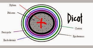
a) Monocot root &
dicot root
dicot root
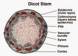
b) Ronocot stem &
dicot stem
dicot stem

Diagram
given in ncert book.
given in ncert book.
Ans
– a) i) Cortex in monocot roots are wider
– a) i) Cortex in monocot roots are wider
ii)In monocot roots larger no of vascular
bundles are present.
bundles are present.
iii) Pith is present in monocot roots.
iv) In monocot root there is rounded
vascular boundless & polygonal in dicot root.
vascular boundless & polygonal in dicot root.
Anms
– b) i) In dicot stem hair is presence & absence in monocot stem.
– b) i) In dicot stem hair is presence & absence in monocot stem.
ii) In dicot stem collenchymatous hypodcermis
is present & sclenenchymatous hypodermis is present in mono cot stem.
is present & sclenenchymatous hypodermis is present in mono cot stem.
iii) Vascular bundlesw are open in
monocot & closed in dicot.
monocot & closed in dicot.
5, Cut a transuerse section of young stem of a
plant from your school garden & obscruit under the microscope. How would
you ascertain wheather it is monocot stem or dicot stem give reason.
plant from your school garden & obscruit under the microscope. How would
you ascertain wheather it is monocot stem or dicot stem give reason.
If the section is of dicot stem if it has
concentric arrangement of ground tissues, open vascular bundles arranged in
polygon vessels.
concentric arrangement of ground tissues, open vascular bundles arranged in
polygon vessels.
If the section is of mono cot stem the
ground tissues are undifferentiated, vascellar bundles are closed.
ground tissues are undifferentiated, vascellar bundles are closed.
6. Why are xylem &
phloem called comples tionves?
phloem called comples tionves?
A complex tissue is one which contains more
than two types of cells which perform common function.
than two types of cells which perform common function.
As xylem is formed of 4 types of cells –
Tracheids, vessels, xylem parenchymja &
xylem fibres which all help in conduction of sap.
xylem fibres which all help in conduction of sap.
Phloem is formed by 4 types of cells –
sieve companion cells, phloem parenchyma & phloem fibres . they help in
conduction of food – to directional
sieve companion cells, phloem parenchyma & phloem fibres . they help in
conduction of food – to directional
7. What is stomatal apparatus ? Explain the
structure of stomata with labelled diagram.
structure of stomata with labelled diagram.
A pair of guard cells with or without
surrounding subsidiary cells that functions as a value to open or close a
stomatal pore forf gaseous exchange & transpirations is called stomatal
appa – ratun.
surrounding subsidiary cells that functions as a value to open or close a
stomatal pore forf gaseous exchange & transpirations is called stomatal
appa – ratun.
The guard cells contains choloropp[last
& small voceloles which are thick – walled in the area opf contact &
thin walked else where. When the guard cells swellep due to on do osmosis the
thin – walled sides expand. The thick walls of the two guarfd cells swell up[
due to endo-osmosis & the thin wall side expands.
& small voceloles which are thick – walled in the area opf contact &
thin walked else where. When the guard cells swellep due to on do osmosis the
thin – walled sides expand. The thick walls of the two guarfd cells swell up[
due to endo-osmosis & the thin wall side expands.
8. Name the three baris tissue systems in the
following flantgs. Give the tissue name under each system.
following flantgs. Give the tissue name under each system.
3
tissues
tissues
1.Epidermal tissue system
2. Ground tissue System
3.Vascular tissue system
Epidermal tissue system
– Epidermis & epidermal appendages. It consist of epidermal & guard
cells. It includes root tairs, stem hairs etc.
– Epidermis & epidermal appendages. It consist of epidermal & guard
cells. It includes root tairs, stem hairs etc.
Ground tissue system –
Hypodermis, corten endodermis, pericycle, pith & medullary rays.
Hypodermis, corten endodermis, pericycle, pith & medullary rays.
Vascular tissue system
– Vascular bundles, phloem, xylem & vascularf cambium.
– Vascular bundles, phloem, xylem & vascularf cambium.
10. What is periderm? How periderm
formation does takes place in dicot stem?
formation does takes place in dicot stem?
It is formed towards the surface of stems &
roots having phellem, phellogm & phelloderm.
roots having phellem, phellogm & phelloderm.
Phellogen or cork
cambium – It develops in a subepiderminal layer in stem & from pericycle in
roots & undergoes bipolar divisions. The all of the outer side undergoes
deposition of tannins & death of cellular contents. The outer tissue of
dead subsidiary cell is called phellem.
cambium – It develops in a subepiderminal layer in stem & from pericycle in
roots & undergoes bipolar divisions. The all of the outer side undergoes
deposition of tannins & death of cellular contents. The outer tissue of
dead subsidiary cell is called phellem.
If the answers or the article helped
you and you liked the site, please support us to avail you more quality study
materials and solutions .Donate us to support us.
you and you liked the site, please support us to avail you more quality study
materials and solutions .Donate us to support us.